🎯Electroencephalography: Brain activity recording
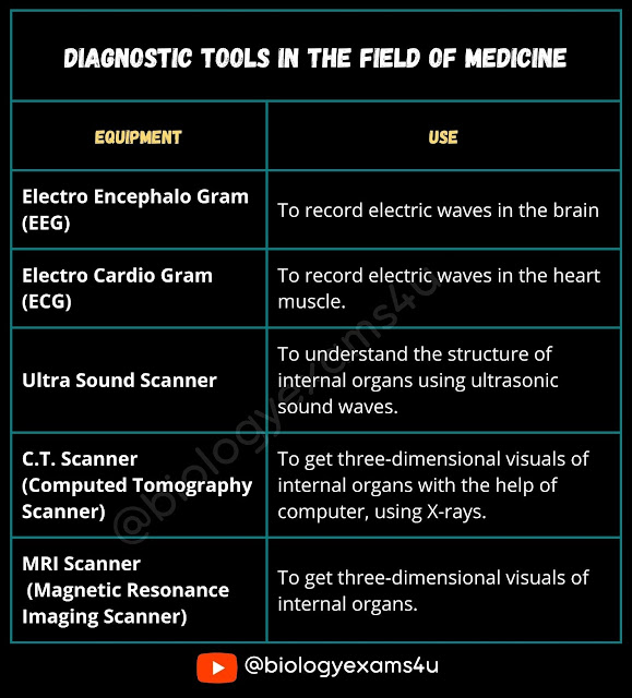
An electroencephalogram (EEG) is a test that measures electrical activity in the brain using small, metal discs (electrodes) attached to the scalp. Brain cells communicate via electrical impulses and are active all the time, even during asleep. This activity shows up as wavy lines on an EEG recording. An EEG is one of the main diagnostic tests for epilepsy. An EEG can also play a role in diagnosing other brain disorders.
🎯Electro encephalogram: Measure the fluctation of dipole voltage in neurons of brain.
🎯Magnetic Resonanace Imaging (MRI) : It is a non-invasive imaging technology that produces three dimensional detailed anatomical images. It is often used for disease detection, diagnosis, and treatment monitoring. It is based on sophisticated technology that excites and detects the change in the direction of the rotational axis of protons found in the water that makes up living tissues.
🎯Computed Axial Tomography: Computed tomography (CT), sometimes called "computerized tomography" or "computed axial tomography" (CAT), is a noninvasive medical examination or procedure that uses specialized X-ray equipment to produce cross-sectional images of the body. Each cross-sectional image represents a “slice” of the person being imaged, like the slices in a loaf of bread. These cross-sectional images are used for a variety of diagnostic and therapeutic purposes.
🎯FMRI (Functional Magnetic Resonance Imaging):Detects changes in blood oxygenation and flow due to neural activity.
🎯Position Emission Tomography : Uses short lived radioacive material to map functional processes in brain.
CT scans can be performed on every region of the body for a variety of reasons (e.g., diagnostic, treatment planning, interventional, or screening)
🎯Single Neuron Recording: There are many techniques available to record brain activity including electroencephalography (EEG), magnetoencephalography (MEG), and functional magnetic resonance imaging (fMRI)—but these do not allow for single-neuron resolution.Neurons are the basic functional units in the brain; they transmit information through the body using electrical signals called action potentials. Currently, single-unit recordings provide the most precise recordings from a single neuron. A single unit is defined as a single, firing neuron whose spike potentials are distinctly isolated by a recording microelectrode. Single neuron recording has revealed the function of many brain areas in terms of stimulus and response.
🎯Patch clamp recording: The patch clamp method is a refined electrophysiological technique that can directly measure the membrane potential and/or the amount of current passing across the cell membrane. The patch clamp technique is also incredibly versatile and can be used in a variety of different configurations to study a range of properties, from spontaneous cell firing activity in native tissue to the activation and/or deactivation kinetics of individual channels expressed in recombinant cell lines.
Question : Given below are spatial and temporal techniques (Column I) used to detect brain activity (Column II)
1. A-i, B-ii, C-iii, D-iv
2. A-iii, B-i, C-iv, D-ii
3. A-iv, B-iii, C-ii, D-i
4. A-ii, B-iv, C-i, D-iii
Answer: 2. A-iii, B-i, C-iv, D-ii
Tags:
methods in biology


